Vaginal Cancer

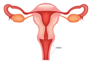
The vagina is a circular, tubular female organ that allows the internal genital organs to open to the external genital organs. It allows penetration of the penis during sexual intercourse.
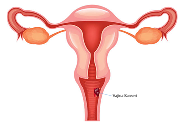
Vaginal cancer is a type of cancer that develops in the vaginal tissue. It usually occurs as a result of other cancers gripping the vagina. Primary cancers of the vagina, on the other hand, are very, very rare.
 Signs(Symptoms)
Early stage vaginal cancers usually do not give any signs. As the cancer progresses, the following findings may be seen.
- Abnormal vaginal bleeding; after intercourse or during menopause.< / li>
- Watery vaginal discharge
- Swelling in the vagina and mass coming to the hand
- Painful Urination
- Frequent urination
- Constipation
- Pelvic pain
When Should I Go to the Doctor?
If the above-mentioned symptoms have become persistent and concern you, I recommend making an appointment with your doctor. But let's not forget, it is the most rational way to have your regular gynecological examinations.
Reasons
Mutations that develop in the DNA of cells located in the vagina lead to the transformation of healthy cells into abnormal cells. Normal cells grow, multiply, and eventually die. Cancerous abnormal cells, on the other hand, grow, multiply uncontrollably and become immortal. This abnormal collection of cells forms a mass and it is called a tumor. Just as cancer cells can invade healthy tissues around them, they can also spread to distant tissues (metastasis).
Types of Vaginal Cancer
- Squamous cell cancer:Develops from flat cells located in the vaginal wall. It is the most common among the primary cancers of the vagina. Decapitation.< / li>
- Adenocarcinoma:Develops from the glandular cells of the vagina.< / li>
- Malignant Melanoma:Develops from pigment-producing cells (melanocytes) of the vagina.< / li>
- Sarcoma:Develops from connective tissue or November muscle cells in the vaginal tissue.< / li>
Risk Factors
- Advanced age:They are usually seen after the age of 60.< / li>
- Vaginal Intraepithelial Neoplasia (VAIN): Problematic cells that have not yet become cancerous in the vagina are called VAIN. VAIN is often caused by the HPV virus, which is a sexually transmitted infection. < / li>
- Diethylstilbestrol (DES) exposure: Women exposed to DES, which was used as a preventive drug for miscarriage in the 1950s, had an increased risk of vaginal cancer in girls.< / li>
- Multiple sexual partners
- Sexual intercourse at an early age
- Smoking
- HIV infection
Protection
There is no 100% way to prevent vaginal cancer. But there are some factors that are known to reduce the risk of vaginal cancer.
- Regular gynecological examination and Smear tests
- HPV vaccines
- Not to smoke
Diagnosis
-
Pelvic examination:Your doctor first observes the outer part of your genital area (vulva). It places a special device called a speculum inside your vagina and evaluates the inside of the vagina and the cervix (cervix). Then, he inserts two fingers into the vagina to check for a mass in the vagina and cervix. Some structures that are invisible to the eye are easier to feel with the fingertip. Again, during the procedure, your doctor presses on your stomach with the other hand, compressing the uterus and ovaries from both sides and tries to find out if there is a mass coming into the hand.
< / li>
- Colposcopic biopsy: Pathological evaluation is essential to confirm the diagnosis of cancer. The suspicious area in the vagina is examined with a special device called colposcopy, which allows it to be observed better by enlarging the tissues, and a biopsy is performed from this suspicious area. < / li>
Staging
If your doctor decides that you have vaginal cancer, he will want to conduct further tests to see if the cancer has spread to other organs. The stage of cancer is the most important factor that determines the treatment.
- Imaging Techniques: Advanced tests such as tomography, MRI, PET Sa-CT can be used to determine the spread of cancer.< / li>
- Imaging of the Bladder and Intestines: If necessary, an image can be taken from the bladder and large intestine with a camera. < / li>
Treatment
The treatment of vaginal cancer depends on various factors. The stage of the cancer, other diseases you have, and your preferences will affect the treatment decision. Treatment options are created with surgery or radiation therapy.
Surgery
- Removal of small tumors and lesions: Superficial lesions located in the vagina can be removed with some healthy surgical limits. < / li>
- Vaginectomy: Part of the vagina (partial vaginectomy) or all of the vagina (radical vaginectomy) can be removed so that the entire cancer can be cleared. During this procedure, the uterus, ovaries and regional lymph nodes can also be removed. < / li>
- Pelvic exenteration: If the vaginal cancer has invaded the surrounding tissues, this radical surgical procedure may be necessary. During the procedure, the vagina, vulva, uterus, ovaries, bladder and large intestine are radically removed in one piece. Stomata are opened to the abdominal wall for urinary and defecation functions.< / li>
Radiation Therapy
Radiation is a special type of energy carried by waves and particles. The use of radiation produced in special devices in the treatment of cancer is called radiotherapy or radiation therapy.
It can be used for vaginal cancer in the following ways;
- By external route, that is, by external irradiation therapy (External beam radiation therapy)
- By internal route, i.e. by vaginal irradiation (Brachytherapy)
Summary
In cancer surgery the right surgeon, the right technology and the right pathology are essential. I strongly recommend that you research your surgeon, get opinions from patients he has operated on before, and question the technologies that will be used in your surgery, and where and how the pathological examination will be performed.

 Signs(Symptoms)