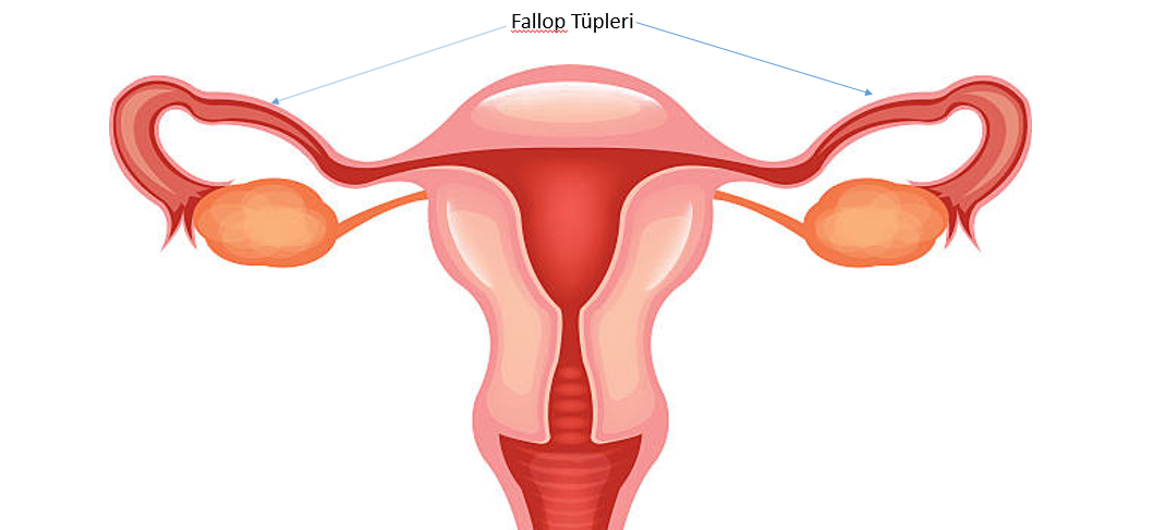
Cancer of the Fallopian Tube (Tuba)
Tubes (tubes) are thin, tubular structures that connect the ovaries to the uterus. Women ovulate once every month during their fertile years. This egg cell is fertilized with sperm in the fallopian tube and passes through the tube to the uterus.

Since the fallopian tube is anatomically very close to the ovary, cancers that develop from these two organs have similarities to each other at the cellular level. For this reason, it is also difficult to determine which organ the cancer started from. Tubal cancer develops from cells that grow uncontrollably, which develop inside the tuba tissue. The treatment of this cancer often requires surgery and chemotherapy.
Symptoms (Signs)
Tubal cancer is also insidious, like ovarian cancer, it often does not give signs when it first develops. When it starts to cause complaints, there are usually delays in its diagnosis because it is confused with the findings of other diseases. The following findings can be seen;
- Bleeding unrelated to menstruation
- Bloody watery discharge
- Abdominal pain (usually in the form of spasms)
- Abdominal swelling
When Should I Go to the Doctor?
If the above-mentioned symptoms have become persistent and concern you, I recommend making an appointment with your doctor. But let's not forget, it is the most rational way to have your regular gynecological examinations.
Reasons
Mutations that develop in the DNA of cells located in tubes lead to the transformation of healthy cells into abnormal cells. Normal cells grow, multiply, and eventually die. Cancerous abnormal cells, on the other hand, grow, multiply uncontrollably and become immortal. This abnormal collection of cells forms a mass and it is called a tumor. Just as cancer cells can invade healthy tissues around them, they can also spread to distant tissues (metastasis).
Risk Factors
- Advanced age:As age progresses, the incidence of tubal cancer increases. They are more common in older women.< / li>
- Hereditary genetic changes: A small proportion of tubal cancers develop due to genetic mutations passed on from parents. These genes are BRCA 1 and BRCA 2 genes. The risk of breast cancer has also increased in women who have inherited these genes.< / li>
- Having a family history of ovarian or tubal cancer: If there are women with ovarian or tubal cancer in your family, it means that you also have an increased risk of this cancer.< / li>
- Obesity
- Hormone replacement therapy after menopause: Synthetic hormones used to treat complaints that develop due to menopause may increase the risk of tubal cancer. < / li>
- Endometriosis: Endometriosis, a disease that occurs when the intrauterine layer settles in places where it should not be in the body, may increase the risk of tubal cancer.< / li>
- Menstruation for many years: Starting menstruation at an early age or entering menopause late.< / li>
- Having never been pregnant
Protection
There is no 100% way to prevent tubal cancer. But there are some factors that are known to reduce the risk of tubal cancer.
- Using birth control pills: In addition to providing high levels of protection, these pills also reduce the risk of tubal cancer. But they may not be suitable for every patient due to side effects. You can discuss with your doctor whether birth control pills are suitable for you.< / li>
- Discuss your personal risk factors with your doctor: If there are patients with ovarian or tubal cancer in your family, you should share this with your doctor. In such a case, you may be referred for genetic counseling. If it is determined by genetic tests that you carry risky gene mutations for ovarian and tubal cancer, it may be recommended to surgically remove your ovaries and tubes before the cancer develops. < / li>
Diagnosis
- Pelvic examination: Your doctor first observes the outer part of your genital area (vulva). It places a special device called a speculum inside your vagina and evaluates the inside of the vagina and the cervix (cervix). Then, he inserts two fingers into the vagina to check for a mass in the vagina and cervix. Some structures that are invisible to the eye are easier to feel with the fingertip. Again, during the procedure, your doctor presses on your abdomen with the other hand, compresses the uterus and adnexes from both sides and tries to find out if there is a mass coming to the hand.< / li>
- Imaging tests: The structure of the ovaries and tubes can be evaluated using imaging techniques such as ultrasound, tomography, MRI. Ultrasound performed vaginally is like the hands and feet of gynecologists and gives very valuable findings in the evaluation of tubes. < / li>
- Blood tests: Your doctor may order blood tests to assess your overall health and tumor markers that may indicate an increase in tubal cancer. < / li>
- Diagnostic laparoscopy: Sometimes in between cases, in order to Decipher the diagnosis, both images are taken and tissue and fluid samples can be taken by entering the abdomen through small holes. < / li>
- Genetic Tests: Your doctor may order genetic tests to determine the presence of hereditary ovarian or tubal cancer. These tests can help plan your treatment, as well as help determine the risk factors of your female relatives and girls.< / li>
Treatment
The treatment of tubal cancer is usually planned by combining surgery and chemotherapy. Different treatments may also be added in certain cases.
Surgery
The main goal of surgery is to remove cancerous tissues and determine the stage of cancer. The stage of the disease also determines the decision on chemotherapy after surgery.
- Salpingo-oophorectomy: In early stage tubal cancers, only one ovary and the accompanying tube can be removed if there are no splashes in other organs. In this operation, the patient's fertility is preserved. In addition, fatty tissue and lymph nodes called omentum in the abdomen can also be removed.< / li>
- Removal of the uterus and each adnexa: In patients who do not want fertility, the uterus, ovaries, tubes, lymph nodes and fatty tissue called the omentum in the abdomen can be removed.< / li>
- Advanced stage cancer surgery: If the cancer is widespread in the abdomen, a difficult operation can be performed in which all the tumor tissues are removed. Sometimes chemotherapy is given to the patient before and the operation is performed afterwards.< / li>
Chemotherapy
Special drugs are used in chemotherapy that destroy fast-growing cells. Cancer cells are also affected by chemotherapy because they grow fast. These medications can be given intravenously or orally.
Chemotherapy is usually used to destroy tumor cells that are invisible to the eye after surgery. In some cases, they can also be used before surgery.
Summary
In cancer surgery the right surgeon, the right technology and the right pathology are essential. I strongly recommend that you research your surgeon, get opinions from patients he has operated on before, and question the technologies that will be used in your surgery, and where and how the pathological examination will be performed.
